|
Go to the MDMA (Ecstasy) Drug Summary Page
|
Ecstasy. A not so bright idea.
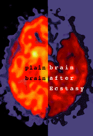
The brains scans on the front of this card show the sharp difference in human brain function for an individual who has never used drugs and one who used the club drug Ecstasy (XTC, MDMA, Adam, etc.) many times, but had not used any drugs for at least 3 weeks before having the scan. The left, bright reddish half shows active serotonin sites in the brain. Serotonin is a critical neurochemical that regulates mood, emotion, learning, memory, sleep, pain. The dark sections in the right half are serotonin sites that are not present even after 3 weeks without any drugs. In addition to these changes in serotonin sites, scientsts have found that Ecstasy injures serotonin neurons. Although these can regrow, they don't grow back normally and might not grow back in the right location.
This image is a composite of scans from the paper referenced below; each half is from the same section of the brain. The graphic and text above are taken from a free postcard NIDA distributes nationwide.
McCann, U.D., Szabo, Z., Scheffel, U., Dannals, R.F., and Ricaurte, G. A. Positron emission tomographic evidence of toxic effect of MDMA ("Ecstasy") on brain serotinin neurons in human beings. The Lancet, Volume 352, October 31, 1998.
|